Shoulder Conditions
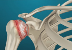
Arthritis of the Shoulder
The term arthritis literally means inflammation of a joint but is generally used to describe any condition in which there is damage to the cartilage. Damage of the cartilage in the shoulder joint causes shoulder arthritis. Inflammation is the body's natural response to injury. The warning signs that inflammation presents are redness, swelling, heat, and pain.
Shoulder Instability
Shoulder instability is a chronic condition that causes frequent dislocation of the shoulder joint. A dislocation occurs when the end of the humerus (ball portion) partially or completely dislocates from the glenoid (socket portion) of the shoulder.
Rotator Cuff Tear
A rotator cuff is a group of tendons in the shoulder joint that provides support and enables a wide range of motion. A major injury to these tendons may result in rotator cuff tears. It is one of the most common causes of shoulder pain in middle-aged and older individuals.
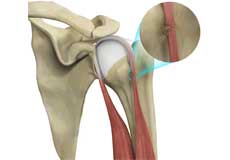
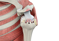
Proximal Biceps Tendon Rupture
The biceps muscle is the muscle of the upper arm which is necessary for the movement of the shoulder and elbow. It is made of a ‘short head’ and a ‘long head’ which function together. These are connected to the shoulder joint by two tendons called the proximal biceps tendons and to the elbow joint by a single distal biceps tendon.
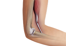
Distal Bicep Tears
The biceps is a large muscle located in the front of your upper arm and runs from the shoulder to the elbow joint. It is attached to the bones of the shoulder and elbow by tendons. The distal biceps is the area where the biceps is attached to the forearm bone in the elbow.
SLAP Tears
The term SLAP (superior –labrum anterior-posterior) lesion or SLAP tear refers to an injury of the superior labrum of the shoulder. The most common causes include falling on an outstretched arm, repetitive overhead actions such as throwing and lifting a heavy object. Overhead and contact sports may put you at a greater risk of developing SLAP tears.
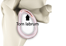
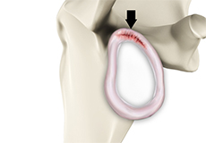
Shoulder Labral Tear
Traumatic injury to the shoulder or overuse of the shoulder (throwing, weightlifting) may cause the labrum to tear. In addition, aging may weaken the labrum leading to injury.
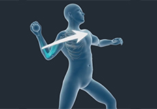
Throwing Injuries of the Shoulder
Throwing injuries of the shoulder are injuries sustained as a result of trauma by athletes during sports activities that involve repetitive overhand motions of the arm as in baseball, American football, volleyball, rugby, tennis, track and field events, etc.
Shoulder Procedures
Rotator Cuff Repair
Rotator cuff repair is a surgery to repair an injured or torn rotator cuff. It is usually performed arthroscopically on an outpatient basis. An arthroscope, a small, fiber-optic instrument consisting of a lens, light source, and video camera. The camera projects images of the inside of the joint onto a large monitor, allowing your doctor to look for any damage, assess the type of injury and repair it.
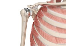
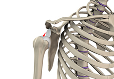
Shoulder Joint Replacement
Total shoulder replacement surgery is performed to relieve symptoms of severe shoulder pain and disability due to arthritis. In this surgery, the damaged articulating parts of the shoulder joint are removed and replaced with artificial prostheses. Replacement of both the humeral head and the socket is called a total shoulder replacement.
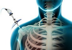
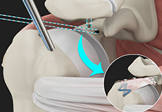
Shoulder Arthroscopy
Arthroscopy is a minimally invasive diagnostic and surgical procedure performed for joint problems. Shoulder arthroscopy is performed using a pencil-sized instrument called an arthroscope. The arthroscope consists of a light system and camera that projects images of the surgical site onto a computer screen for your surgeon to clearly view.
Shoulder Stabilization
Shoulder stabilization surgery is performed to improve stability and function to the shoulder joint and prevent recurrent dislocations. It can be performed arthroscopically, depending on your particular condition, with much smaller incisions. Arthroscopic stabilization is a surgical procedure to treat chronic instability of the shoulder joint.
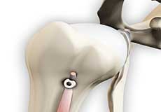
Proximal Biceps Tenodesis
Proximal biceps tenodesis is the surgical reattachment of a torn proximal biceps tendon, which connects the upper part of your biceps muscle to the shoulder.
Pectoralis Major Tears/Repairs
A tear in the pectoralis major muscle occurs when the tendon attaching the muscle to the upper arm bone is damaged as a result of trauma or injury. In the case of a rupture, operative treatment is employed for repairing the torn muscle in active and young patients irrespective of the chronicity of the injury.
Arthroscopic Superior Capsular Reconstruction (SCR)
Superior Capsular Reconstruction is a surgical procedure to repair massive, irreparable rotator cuff tears. The surgery involves reconstruction of the superior capsule of the shoulder joint using an autograft (tissue from the same person) or an allograft (tissue from a donor).
Shoulder Anatomy
The shoulder is the most flexible joint in the body that enables a wide range of movements including forward flexion, abduction, adduction, external rotation, internal rotation, and 360-degree circumduction. Thus, the shoulder joint is considered the most insecure joint of the body, but the support of ligaments, muscles, and tendons function to provide the required stability.
Bones of the Shoulder
The shoulder joint is a ball and socket joint made up of three bones, namely the humerus, scapula, and clavicle.
Humerus
The end of the humerus or upper arm bone forms the ball of the shoulder joint. An irregular shallow cavity in the scapula called the glenoid cavity forms the socket for the head of the humerus to fit in. The two bones together form the glenohumeral joint, which is the main joint of the shoulder.
Scapula and Clavicle
The scapula is a flat triangular-shaped bone that forms the shoulder blade. It serves as the site of attachment for most of the muscles that provide movement and stability to the joint. The scapula has four bony processes - acromion, spine, coracoid and glenoid cavity. The acromion and coracoid process serve as places for attachment of the ligaments and tendons.
The clavicle bone or collarbone is an S-shaped bone that connects the scapula to the sternum or breastbone. It forms two joints: the acromioclavicular joint, where it articulates with the acromion process of the scapula and the sternoclavicular joint where it articulates with the sternum or breast bone. The clavicle also forms a protective covering for important nerves and blood vessels that pass under it from the spine to the arms.
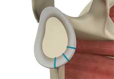
Soft Tissues of the Shoulder
The ends of all articulating bones are covered by smooth tissue called articular cartilage, which allows the bones to slide over each other without friction, enabling smooth movement. Articular cartilage reduces pressure and acts as a shock absorber during movement of the shoulder bones. Extra stability to the glenohumeral joint is provided by the glenoid labrum, a ring of fibrous cartilage that surrounds the glenoid cavity. The glenoid labrum increases the depth and surface area of the glenoid cavity to provide a more secure fit for the half-spherical head of the humerus.
Ligaments of the Shoulder
Ligaments are thick strands of fibers that connect one bone to another. The ligaments of the shoulder joint include:
Coracoclavicular ligaments: These ligaments connect the collarbone to the shoulder blade at the coracoid process.
Acromioclavicular ligament: This connects the collarbone to the shoulder blade at the acromion process.
Coracoacromial ligament: It connects the acromion process to the coracoid process.
Glenohumeral ligaments: A group of 3 ligaments that form a capsule around the shoulder joint and connect the head of the arm bone to the glenoid cavity of the shoulder blade. The capsule forms a watertight sac around the joint. Glenohumeral ligaments play a very important role in providing stability to the otherwise unstable shoulder joint by preventing dislocation.
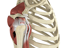
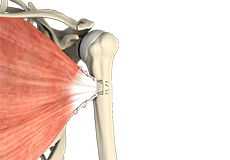
Muscles of the Shoulder
The rotator cuff is the main group of muscles in the shoulder joint and is comprised of 4 muscles. The rotator cuff forms a sleeve around the humeral head and glenoid cavity, providing additional stability to the shoulder joint while enabling a wide range of mobility. The deltoid muscle forms the outer layer of the rotator cuff and is the largest and strongest muscle of the shoulder joint.
Tendons of the Shoulder
Tendons are strong tissues that join muscle to bone allowing the muscle to control the movement of the bone or joint. Two important groups of tendons in the shoulder joint are the biceps tendons and rotator cuff tendons.
Bicep tendons are the two tendons that join the bicep muscle of the upper arm to the shoulder. They are referred to as the long head and short head of the bicep.
Rotator cuff tendons are a group of four tendons that join the head of the humerus to the deeper muscles of the rotator cuff. These tendons provide more stability and mobility to the shoulder joint.
Nerves of the Shoulder
Nerves carry messages from the brain to muscles to direct movement (motor nerves) and send information about different sensations such as touch, temperature, and pain from the muscles back to the brain (sensory nerves). The nerves of the arm pass through the shoulder joint from the neck. These nerves form a bundle at the region of the shoulder called the brachial plexus. The main nerves of the brachial plexus are the musculocutaneous, axillary, radial, ulnar and median nerves.
Blood vessels of the Shoulder
Blood vessels travel along with the nerves to supply blood to the arms. Oxygenated blood is supplied to the shoulder region by the subclavian artery that runs below the collarbone. As it enters the region of the armpit, it is called the axillary artery and further down the arm, it is called the brachial artery.
The main veins carrying de-oxygenated blood back to the heart for purification include:
- Axillary vein: This vein drains into the subclavian vein.
- Cephalic vein: This vein is found in the upper arm and branches at the elbow into the forearm region. It drains into the axillary vein.
- Basilic vein: This vein runs opposite the cephalic vein, near the triceps muscle. It drains into the axillary vein.